Scientists have long studied the neuromuscular junction, or NMJ, which bridges nerves to muscle fibers. This bridge carries signals that cause muscle contractions, making it a linchpin for movement – and the loss of it.
Now, after decades of NMJ research, a team led by scientists at the Robert J. and Nancy D. Carney Institute for Brain Science have uncovered surprising new insights about how it’s built and how it works. In The Journal of Neuroscience, they share their discovery of a second, wholly distinct role for muscle-specific kinase, or MuSK, a protein found at the NMJ. Researchers found that MuSK assembles sodium channels, which amplify muscle contraction signals from the neurotransmitter acetylcholine.
They discovered this role for MuSK when they removed a critical piece of the protein in mice and disrupted the amplifier – which weakened the contractions.
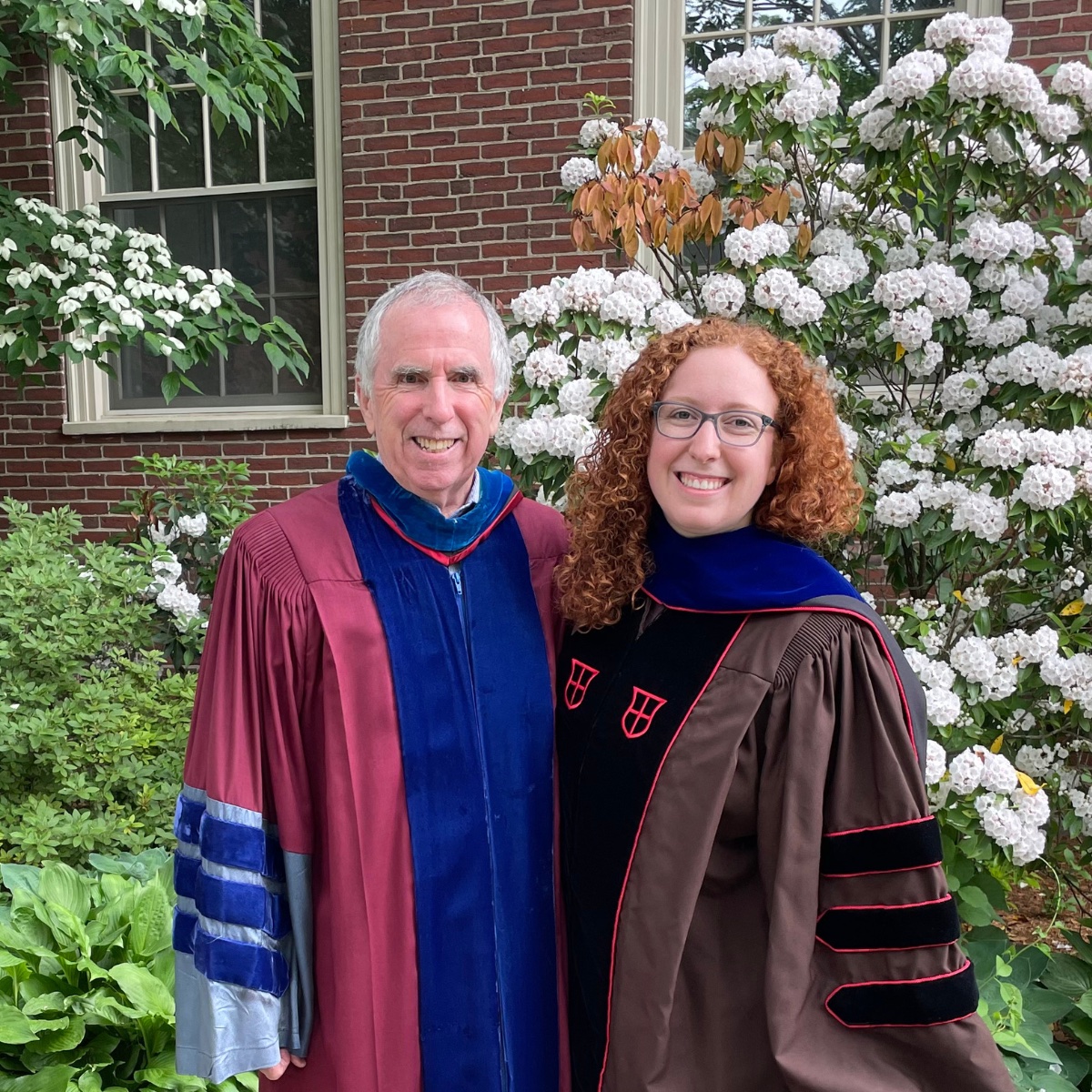
“This was a complete surprise,” said Lauren Fish, a University of Michigan postdoctoral research fellow and first author on the new publication, who made the discovery as a graduate student in the lab of Carney’s Justin Fallon. “Our finding really recasts how we, as researchers, think about the neuromuscular junction and the movement it makes possible.”

A Brown professor of neuroscience, Fallon has spent years studying the NMJ and its essential role in transmitting electrical impulses from the nervous system to the muscles so the body can breathe, swallow, and perform voluntary movements like lifting and walking. Fallon said researchers have long known that MuSK gathers and assembles acetylcholine receptors – which help the body pick up the contraction signals. What’s new, after two generations of study, is that MuSK also assembles sodium channels, which amplify those signals.
“This means we have a new target for drugs to treat conditions where you see muscle weakness or wasting, like spinal muscular atrophy, amyotrophic lateral sclerosis, even Duchenne muscular dystrophy,” Fallon said.
Fish also noted the potential implications for sarcopenia – the gradual loss of muscle mass, strength, and function that occurs with aging. According to the National Institutes of Health, people lose about 50% of their muscle mass by the time they’re 80 years old. This can make everyday activities like getting out of a chair difficult and increase the likelihood of falls.
Fish and Fallon are working with international collaborators to take their findings to the next level, work they hope will provide evidence of a direct connection between sodium channels and sarcopenia.
Fish was a PhD student in Fallon’s lab studying the NMJ alongside graduate students Diego Jaime and Laura Madigan and undergraduate Madison Ewing. Graduate student Atilgan Yilmaz had already discovered in cell cultures that MuSK did more than organize acetylcholine receptors. To study this, Jaime used the CRISPR gene editing tool to make a mutant mouse that lacked a key part of the MuSK protein. Using these mice, the trio found new cellular roles for MuSK – but didn’t have the tools to study signaling from nerve to muscle at the NMJ.
Their options were limited. It was 2021 and the COVID-19 pandemic was raging. Many labs were shuttered. In a virtual session at the Society for Neuroscience annual meeting, Fish was discussing the intriguing new mouse model and wished there was a collaborator who could study it. Kelly Rich, a researcher working at The Ohio State University in the lab of David Arnold, MD, piped up.
“We can do this,” Rich said.
They sent mutant mice to Arnold’s lab, where researchers used sensors on the mouse’s skin to record electrical impulses from the muscle in response to signals from the nerve. Those impulses from the muscle were smaller than usual. Then they looked deeper, putting electrodes into individual muscle fibers, and found the muscle’s responses to nerve stimulation were erratic and unreliable.
But they still didn’t know if the problem was at the synapse – and what caused it. To get that information, they would need to study the NMJ itself.
The mice were then sent to the Wright State University lab of Mark Rich, MD, PhD, where researchers stimulated the nerve and recorded the response from the NMJ directly. Tests showed that the nerve was releasing normal amounts of acetylcholine – and the acetylcholine receptors were picking it up. Yet they knew that in mice, the muscle contractions were weak. Chengjie Xi, a former graduate student in the Fallon lab, found that the MuSK mutant mice had reduced levels of sodium channels at the synapse. This discovery made all the findings fall into place: Alter the protein, break the amplifier, weaken the contractions.
“We learned that MuSK assembles sodium channels, which are a key part of the cell signal cascade that causes muscles to contract,” Fish said. “Amplification is hugely important for healthy nerve to muscle signaling and for normal muscle movement.”
The research was supported by a Carney Institute for Brain Science Graduate Award, a Brown University Undergraduate Research and Teaching Award, and an Emerging Areas of New Science DEANS Award from the Division of Biology and Medicine at Brown University.
Federal grant support came from the National Institute on Aging, (2T32 AG041688, 1F99 AG079815-01, R01 AG078129, R01 AG06775, R21 AG073743, R41 AG073144); the National Institute of Neurological Disorders and Stroke (2T32 MH020068, U01 NS064295, R21 NS112743), the National Institute of General Medical Sciences (4R25 GM083270, R01 GM068118); the National Institute of Arthritis and Musculoskeletal and Skin Diseases (R01 AR074985, R01 AR074985) and the NIH Office of the Director (1S10 OD025181).
Further support came from the Cleft Palate Foundation, ALS Finding a Cure and an Ohio State University Presidential Fellowship.